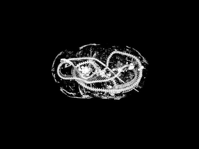
Microscopic scans revealed a mummified snake from ancient Egypt was killed with a whipping action (Swansea University)
Sign up to our free weekly IndyTech newsletter delivered straight to your inbox
Sign up to our free IndyTech newsletter
Researchers have “digitally unwrapped” mummified animals from ancient Egypt, revealing previously undiscovered details about ritualistic mummification.
Analysis of the 2,000-year-old animals – which include a bird, a kitten and a snake – was done using a technique that achieves a resolution 100-times higher than a medical CT scan.
“Using micro CT we can effectively carry out a post-mortem on these animals, more than 2,000 years after they died in ancient Egypt,” said Professor Richard Johnston from Swansea University, who led the research.
“We were able to piece together new evidence of how they lived and died, revealing the conditions they were kept in, and possible causes of death.
“These are the very latest scientific imaging techniques. Our work shows how the hi-tech tools of today can shed new light on the distant past.”
Ancient Egyptians mummified humans as well as pets and other animals, including hawks, ibis and crocodiles. It is estimated that up to 70 million animal mummies were embalmed by temple priests.
The microscopic scans indicated that the kitten had been strangled, while the snake had been killed by a whipping action.
The researchers believe this constitutes the first ever evidence that complex ritualistic behaviour was applied to a snake.
Gadget and tech news: In pictures
Show all 25
Micro CT scans offer significant benefits over methods previously used to examine mummies. Standard X-Rays only provide 2-dimensional images, while medical CT scans only produce low-resolution images.
The analysis technique is typically used by engineers to scan jet engine parts, composites and other industrial materials.
Once scanned, the images can be either 3D printed or placed into virtual reality in order to conduct further analysis.
“This collaboration between engineers, archaeologists, biologists, and Egyptologists shows the value of researchers from different subjects working together,” said Dr Carolyn Graves-Brown from the Egypt Centre at Swansea University.
“Our findings have uncovered new insights into animal mummification, religion and human-animal relationships in ancient Egypt.”
The seven-year research project, which involved collaboration between experts at Swansea, Cardiff and Leicester universities, was published in the journal Scientific Reports.
Join our commenting forum
Join thought-provoking conversations, follow other Independent readers and see their replies

Join our commenting forum
Join thought-provoking conversations, follow other Independent readers and see their replies